

AgenticHealth
On-premise clinical platform with local LLMs, RAG on FHIR/DICOM data, diagnostic support, remote follow-up. Architecture designed for the MDR pathway.
Discover AgenticHealth →
Digital Health
Medical software compliant with CE and MDR standards. Clinical decision support systems and AI integration in clinical workflows.
Discover →
Research & Development
Applied research and prototyping, from biomedical imaging to AI, in partnership with universities and clinical centres.
Discover →
Open Source
noze’s contribution to the Open Source ecosystem: projects, tools and publications released under permissive licences.
Discover →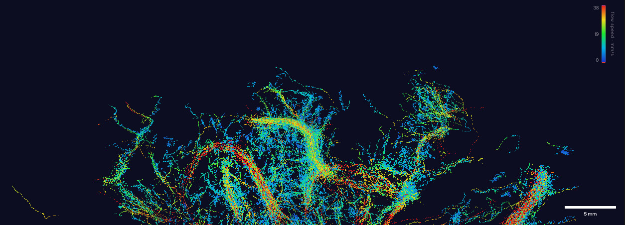
What Aleph Neuro showed
On 24 June 2026 the US lab Aleph (Aleph Neuro) published what it describes as the most detailed vascular image ever obtained of a living human brain, captured with ultrasound through the intact skull. The next day, introducing itself publicly as “a research lab building brain interfaces for the telepathic future”, the company shared the result as “the highest-resolution 3D images of the human brain ever taken from outside the skull”.
According to Aleph this is the world’s first 3D image of ultrasound localization microscopy (ULM) in a human brain through a skull, with a claimed resolution up to 100 times greater volumetrically than comparable CT: sub-millimetre structures and roughly a million independent “voxels” throughout the brain. These are self-reported figures, still to be confirmed by peer review, but the order of magnitude is remarkable for a non-invasive technique with no ionising radiation.
The technique: localization microscopy (ULM)
Conventional ultrasound has a physical limit: it cannot resolve detail smaller than the wavelength of the beam (the diffraction limit). Ultrasound localization microscopy sidesteps that limit with an elegant idea borrowed from optical super-resolution microscopy.
A contrast agent made of microbubbles (sulfur hexafluoride encapsulated in a lipid shell, an agent already FDA-approved and used in contrast-enhanced ultrasound) is injected into the bloodstream. The bubbles are strong ultrasound reflectors. Inject them dilute enough that their echoes don’t overlap, and you can pinpoint the centre of each bubble with a precision finer than the wavelength itself. By tracking millions of these positions as the bubbles flow through the vessels, you reconstruct the microvasculature with detail impossible for conventional ultrasound, down to colouring each segment by flow speed.
Aleph reports an acquisition of about four minutes with continuous contrast infusion, and a pipeline that compresses the raw data to roughly 0.1% of the original volume. The real challenge, through the skull, is exactly this: bone attenuates and distorts ultrasound, which is why 3D transcranial ULM in humans had not been demonstrated until now.
From the $100,000 scanner to the smartphone
There is an enabling factor worth highlighting. Ultrasound hardware, Aleph notes, has gone within a few years from machines costing “$100,000 and requiring a cart full of electronics” to devices “about the price and size of a smartphone”. It is the same trajectory that made computational photography possible: less dedicated hardware, more signal processing and software. When the physics moves into software, reproducibility and access change scale. And this is where the most interesting choice comes in for those of us who work on medical software and open source.
The pipeline is open source (GitHub)
Aleph released the entire pipeline along with the dataset. The repository is public on GitHub: github.com/alephneuro/microbubbles (MIT licence), written mostly in Python, with interactive viewers in JavaScript/HTML. The link cited in the original article (alephneuro/braindump) now redirects to this same repository.
The workflow is organised in three stages, exposed as subcommands:
- Beamforming: from raw ultrasound data (demodulated IQ) to a reconstructed volume in H5 format, with optional GPU acceleration (CUDA).
- Tracking: temporal SVD clutter filtering, 3D sub-voxel localization of the microbubbles, track linking with a Kalman filter and smoothing.
- Viewing: export of compact tracks and in-browser viewers (animated tracks, rotatable 3D volume).
A sample dataset ships alongside the code (about 98 GB, 223 acquisitions), with demodulated IQ data and the beamforming configuration. For researchers, this is the detail that matters more than the headline: not a press release, but code and data that let you reproduce and verify the result, and build on top of it.
Why it matters for the clinic
Cerebral microvasculature is a difficult diagnostic target. Conditions like stroke, Alzheimer’s disease and traumatic brain injury each leave vascular signatures at scales CT and MRI struggle to resolve. Non-invasive imaging, with no radiation, at micrometre resolution and on ever cheaper hardware, opens up screening and monitoring scenarios that are hard to picture today at the bedside. Aleph points to contrast-free neurovascular imaging as the next direction, which would remove even the microbubble infusion.
Let’s be honest about where we are: this is a research result, with figures stated by the company and not yet independently validated. The road to a certified medical device (CE marking, MDR pathway, clinical studies) is long. But the technological base is solid and, above all, it is open.
Our take
What strikes me most is how much the value is shifting into software and AI. When a scanner costs as much as a smartphone and the physics of acquisition becomes signal processing, the clinical differentiator is no longer the machine but the pipeline that extracts meaning from it: reconstruction, sub-voxel localization, tracking and, increasingly, AI models that assist detection and interpretation. Advanced diagnostics, today, is first and foremost a software problem. This is exactly the ground we work on with Digital Health and AgenticHealth: software compliant with CE/MDR, on-premise, with AI built into clinical workflows and the patient’s data kept under control.
And it won’t be an isolated case. Just six days before Aleph, on 18 June 2026, Midjourney announced Midjourney Medical: a 60-second full-body ultrasound scanner, meant to make preventive imaging as accessible as a trip to the spa. Two announcements in the same week, both pushing ultrasound and AI toward non-invasive, affordable, potentially mass-scale diagnostics. We’ll see more and more of this: it is one of the most promising directions of the coming years. What a time to be alive, you might say, and even more so to be able to contribute to this revolution in healthcare.
And a very concrete way to contribute is open source. Releasing the pipeline and dataset under an MIT licence, as Aleph did, makes the difference between an announcement and a shared, verifiable advance: it is the same model that made 3D Slicer a reference platform for medical imaging. Not everything gets released, of course: proprietary hardware, models and sensitive data often stay closed (Midjourney, on this point, is the exact opposite of Aleph). But when code and data are open, research accelerates and results can be reproduced, which is why noze contributes to open source and designs its products to be auditable.
One methodological note holds for all of this: the extraordinary figures (the “world’s first”, the “100 times CT”, the “30% of deaths avoidable”) are for now claims made by the companies, to be confirmed by peer review, and the road to certified medical devices (CE, MDR, clinical studies) is long. But the direction is clear, and it is genuinely exciting to get to work on it.
Sources
- Aleph Neuro: Ultrasound imaging of the brain
- Launch announcement (X / @alephneuro)
- GitHub repository: alephneuro/microbubbles (pipeline + dataset, MIT)
- 3D transcranial ULM in awake mice: protocol and open-source pipeline (Communications Engineering)