
Contents
Digital Health
MDR, FHIR, clinical RAG: AI in healthcare with the data staying inside the organisation’s perimeter.
Discover →
Research and Development
Applied research on AI, imaging and robotics, with clinical and academic partners.
Discover →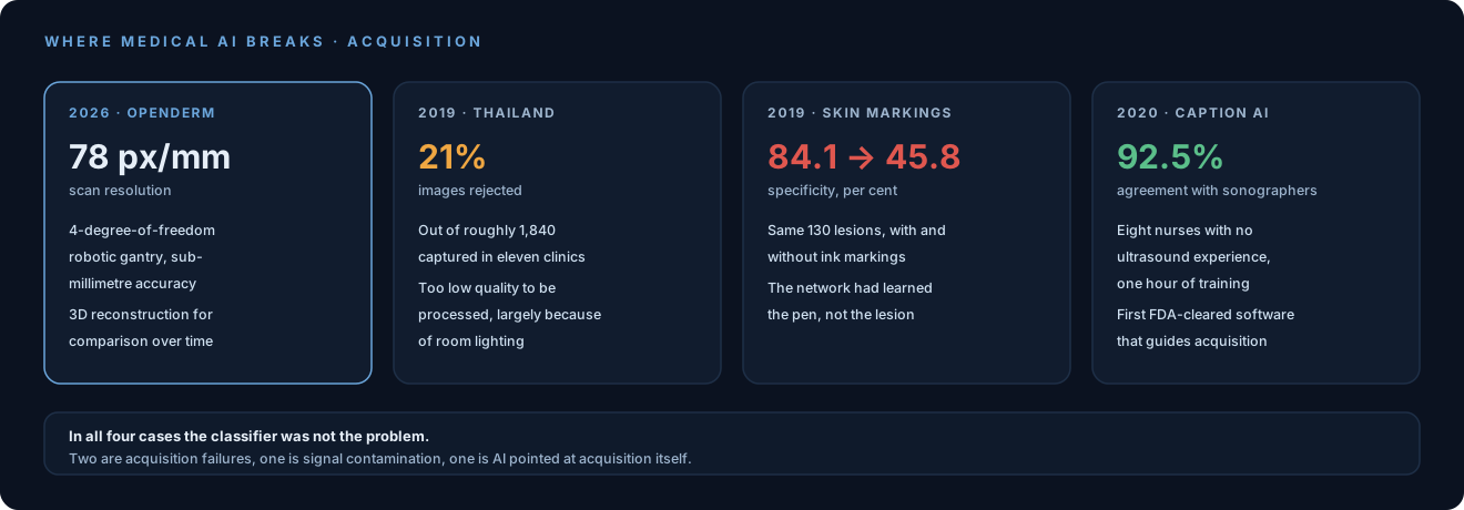
What the post argues
In July 2026 Marion Lepert, described there as a robotics PhD, published “Why Catching Skin Cancer Early Is a Home Robotics Problem” along with OpenDerm, a self-built prototype.
OpenDerm is a 4-degree-of-freedom robotic gantry carrying a camera that scans skin with sub-millimetre positioning accuracy at 78 pixels per millimetre, and reconstructs a 3D map of the skin surface so it can be compared over time. The parts cost around $8,500 and the project is declared open source, with the explicit aim of lowering the barrier to reproducing it.
The thesis is about acquisition. An image has to be acquired well before it can be recognised:
Capturing repeatable, high-resolution images requires moving a camera across the body’s contours while maintaining a consistent distance, viewing angle, focus, and lighting.
Constant distance, angle, focus and lighting while the camera follows the curves of a body. Put that way it is a motion-control specification, not a computer-vision one. And the second argument is even more robotic: an arm knows where it is, so it can geometrically register thousands of overlapping images and compose them into a reconstruction, which a hand holding a phone cannot guarantee.
The numbers it rests on
The post offers two clinical figures to justify the effort. They are its figures, not ours.
First: roughly 70% of melanomas arise as new lesions on previously normal skin, while only about 30% develop from pre-existing moles. This is the figure that undermines the current surveillance model, built entirely on keeping an eye on the moles already there.
Second: five-year survival goes from over 99% for localised melanoma to around 35% once it has spread to distant sites. That is why the time window matters more than the sophistication of the instrument.
Together they say you need to look at all the skin, often, and the same way every time, because what has to be caught is a difference between two scans rather than an anomaly in a single image.
Three cases that say the same thing
This is where the post stops being a personal project and becomes an instance of a category. The medical AI literature has already documented three times that the breaking point is acquisition rather than the classifier.
Thailand, 2019: 21% of images rejected. Google Health took a diabetic retinopathy model with laboratory accuracy comparable to specialists into eleven Thai clinics. In the field evaluation published by Beede and colleagues at CHI 2020, the system rejected 21% of the roughly 1,840 images captured by nurses as too low-quality to process, largely because of room lighting. The model had got nothing wrong: it was not being given an image to work on.
Surgical markings, 2019: specificity collapses. Winkler and colleagues, in JAMA Dermatology, took 130 lesions and passed them to a convolutional network for melanoma recognition. On unmarked lesions the network reached 95.7% sensitivity and 84.1% specificity. On the same lesions with surgical ink markings around them, melanoma probability rose, sensitivity reached 100% and specificity fell to 45.8%, with false positives on benign nevi up by around 40%. Cropping the ink out of the image restored specificity. The authors’ explanation is that in the training data markings appeared mostly next to melanomas: the network had learned the pen.
Caption AI, 2020: AI pointed at acquisition. On 7 February 2020 the FDA authorised, through a De Novo pathway inside the Breakthrough Device programme, the first software that guides the operator in real time while acquiring diagnostic echocardiography images. It does not interpret: it says how to move the probe, with over ninety types of instruction. In the supporting study, eight nurses with no ultrasound experience, after about an hour of training and nine practice scans, produced 240 scans with at least 92.5% agreement with sonographer scans on the main diagnostic endpoints.
The first two are acquisition failures. The third is what happens when AI is pointed at acquisition instead of assuming it solved.
What OpenDerm still lacks
The limits are listed in the post itself, and they cover most of the distance between a prototype and a clinical instrument.
In the current version the patient lies underneath the gantry, and the post itself lists a standing version as a necessary improvement, for safety reasons. Scanning is slow and would move to belt drives instead of ballscrews. Above all, the project does not solve the access problem: a gantry at home is not a realistic prospect, and the post explicitly ties it to the future availability of general-purpose household robots.
Two things the post does not address. First, a device acquiring images for diagnostic purposes falls, in Europe, inside the scope of the medical device regulation: classification and clinical validation are a body of work comparable to the engineering. Second, a 3D map of a person’s entire skin surface, repeated over time, is among the most identifying data there is, and where it gets processed is not an implementation detail.
What we think
Our judgement here is not neutral: we have worked on exactly this problem. From 2019 to 2023, in the ALA project between Gruppo INPECO, a world leader in clinical laboratory automation, and the BioRobotics Institute of Scuola Superiore Sant’Anna, noze acted as AI and cloud software architect. The project ran to 3.5 million euro and thirty researchers, with clinical validation at the dermatology centres of Siena and Livorno.
The hardware. One of the three research lines was the design of an integrated multimodal sensing device with deep learning inference on board: system layout, sub-module design, infrastructure integration. Not a better camera, a machine built to acquire. Alongside RGB images the device collected signals from spectrophotometers, which is information that does not exist to the eye: two lesions can look identical in a photograph and respond differently across bands a camera cannot see. It is the same reasoning as OpenDerm carried along a different axis. The gantry adds geometric repeatability, multimodal sensing adds signal channels, but the move is the same: if acquisition is what limits the system, you build the thing that acquires.
The models. The software line was vertical models for melanoma diagnostic support, not a generic classifier repurposed: architectures built for the case, optimised through neural architecture search techniques (AdaNet, AmoebaNet, NasNet) and hardened with training regimens, data augmentation and data generation. The stated reference point was beating human performance on the task. The third line was end-to-end traceability of biological samples and clinical data along the automation chain, which is the same concern seen from another angle: always knowing where what you are looking at came from.
The app. To get usable images from patients we built a mobile app with guided acquisition: the user does not photograph the lesion however they like, they are walked through doing it in a way comparable to last time. Two photographs taken months apart say something about a lesion only if both were acquired the same way.
Lined up: one research line on the device that acquires, one on the models, one on traceability, plus an app to make photographs comparable. Three of those four workstreams dealt with acquisition, traceability and comparability of the images the classifier reads.
Three observations follow that hold beyond dermatology.
The budget of a medical AI project should go where the risk is, and it is not in the model. On an image classification problem with decent public datasets, a reasonable classifier can be reached with well-established methods. Reaching images acquired repeatably, in a real environment, by untrained people, is where projects fail. If a work plan gives acquisition two weeks out of twenty-four, the plan is wrong.
A model that sees the artefact instead of the disease will not show up in internal validation. The markings case is instructive precisely because the network worked: 95.7% sensitivity, publishable numbers. What was broken lived in the correlation between a pen stroke and a diagnosis, and it surfaced only because somebody thought to vary the acquisition while holding the lesion constant. That is the kind of check that belongs in the test protocol, not left to somebody’s curiosity.
Repeatability is a system requirement, not an operator virtue. The three routes are removing the operator (the gantry), guiding them (Caption AI, our app), or standardising the environment. The fourth, hoping they hold still under good light, is not a route, and the Thai figures say so precisely.
On the idea itself, that early melanoma detection is a positioning problem before it is a vision problem, we agree, and it does not strike us as a minority view among people who have tried to take these systems into a real room. On the gantry at home we are as sceptical as the post itself is. What remains, regardless of whether that prototype goes anywhere, is that opening the hardware of a problem like this is the fastest way to get others to study it.
Sources
- Marion Lepert: Why Catching Skin Cancer Early Is a Home Robotics Problem
- OpenDerm
- Beede et al., A Human-Centered Evaluation of a Deep Learning System Deployed in Clinics for the Detection of Diabetic Retinopathy (CHI 2020)
- Winkler et al., Association Between Surgical Skin Markings in Dermoscopic Images and Diagnostic Performance of a Deep Learning CNN for Melanoma Recognition (JAMA Dermatology, 2019)
- FDA: De Novo authorisation of the first AI software guiding echocardiography acquisition